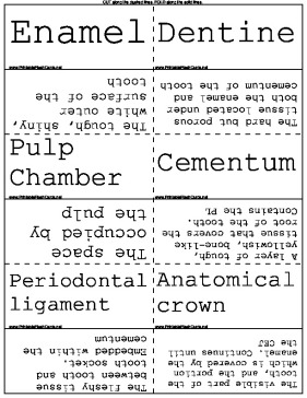

Any dental student will be well served by these detailed dental anatomy flash cards. Study for your next dental exam by learning these words.
There are 44 flash cards in this set (8 pages to print.)
To use:
1. Print out the cards.
2. Cut along the dashed lines.
3. Fold along the solid lines.
Sample flash cards in this set:


| Questions | Answers |
|---|---|
| Enamel | The tough, shiny, white outer surface of the tooth |
| Dentine | The hard but porous tissue located under both the enamel and cementum of the tooth |
| Pulp Chamber | The space occupied by the pulp |
| Cementum | A layer of tough, yellowish, bone-like tissue that covers the root of the tooth. Contains the PL |
| Periodontal ligament | The fleshy tissue between tooth and tooth socket. Embedded within the cementum |
| Anatomical crown | The visible part of the tooth, and the portion which is covered by the enamel. Continues until the CEJ |
| Apical foramen | Natural opening in the root |
| Alveolar bone | The bone which supports the tooth |
| Cortical bone | The dense outer layer of bone |
| Cancellous bone | The sponge like bone found in between two layers of cortical bone |
| Lamina dura | The thin compact bone that lines the alveolar socket (Alveolus) |
| Mandibular vestibule | Space between the teeth and the inner mucosal lining of the lips and cheeks |
| Maxillary tuberosity | Tough hard bumps behind your top back teeth |
| Hamulus | The hard little bumps in the corner of the soft palate |
| Retromolar pad | behind the last lower molars |
| Plane | An imaginary line which divides the body in sections |
| Mid Saggital plane | Divides the patients face into left and right equal sides |
| Frankfurt Plane | Passes through the top of the ear canal and bottom of the eye socket |
| Occlusal plane | The plane passing through the occlusal surfaces of the teeth |
| Coronal plane | A vertical plane at right angles to the mid saggital plane which divides the body into anterior and posterior (front and back) |
| Foramen | Hole or opening in bone |
| Fossa | broad, shallow depression in bone |
| Sinus | Hollow space in bone |
| Canal | Passageway through bone |
| Septum | Bony partition that seperates two spaces |
| Suture | Immovable joint between bones |
| Name the 9 bones of the head | Parietal, Occipital, Frontal, Nasal, Sphenoid, Zygomatic, Maxilla, Mandible, Temporal |
| What is a suture? Name the sutures of the skull | An immovable joint that joins two bones together. Coronal suture, Saggital suture, Median palatine suture, Lamboidal suture |
| What is the Nasal Septum? | A bony wall that seperates the left ad right cavities in the nose |
| What is the nasal cavity? | A large air filled cavity above and behind the nose |
| What is enamel? | The tough, shiny white outer surface of the tooth |
| What is a foramen? | An opening or hole in a bone that permits the passage of nerves and blood vessels |
| What is cortical bone? | The dense outer layer of bone. Appears radiopaque on a dental image |
| What is cancellous bone? | The soft spongy bone located between two layers of dense cortical bone. Appears radiolucent on a dental image |
| What is a process? | A marked prominence or projection of bone |
| Dentine? | The hard but porous tissue located under both the enamel and cementum of the tooth (harder than bone) |
| Pulp Chamber? | The space occupied by pulp |
| Cementum? | A layer of tough, yellowish, bone-like tissue that covers the root. Contains the periodontal ligament |
| Periodontal ligament? | The fleshy tissue between tooth and the tooth socket. The fibers of the PL are embedded within the cementum |
| Anatomical crown? | The visible part of the tooth, the part which is covered by enamel. Goes up to the CEJ |
| Apical foramen? | Natural opening in the root |
| Alveolar bone? | The bone which supports the tooth |
| Lamina Dura? | The thin compact bone that lines the Alveolar socket (Alveolus) |
| Hamulus? | The hard little bumps in the corner of the soft palate |