If you are studying to become an optometrist, you may find these vocabulary cards to be very useful. These cards cover the structure of the eye as well as many disorders involving the eyes.
There are 95 flash cards in this set (16 pages to print.)
To use:
1. Print out the cards.
2. Cut along the dashed lines.
3. Fold along the solid lines.

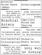
Sample flash cards in this set:


| Questions | Answers |
|---|---|
| Epithelium | First layer of the cornea. asks as the first line of defence against infection and injury |
| Bowman's Membrane | Second layer of the cornea. Acts as an achor to the epithelium. |
| Stroma | Third layer of the cornea. It is the main body of the cornea and contributes rigidity. |
| Descement's Membrane | Fourth layer of the cornea. Contributes rigidity. |
| Endothelium | Fifth layer of the cornea. Serves as pumps to maintain proper fluid balance. |
| Uvea | Iris, Ciliary body and Choroid |
| Axial Length | Length of the from front to back. |
| Adnexa | Orbit, Extra ocular muscles, eyelids, tear-producing and tear-draining lacrimal apparatus. |
| Orbit | Comprising 7 bones that house the globe, extra ocular muscles, blood vessels and nerves. |
| Medial Rectus | Moves eye inward toward the nose |
| Lateral Rectus | Moves the outward toward the temple. |
| Superior Rectus | Moves the eye upward. |
| Inferior Rectus | Moves the eye downward. |
| Superior Oblique | Twists the eye down and in. |
| Inferior Oblique | Twists the eye up and out. |
| Adduction | The movement eye both eye turning in. |
| Abduction | The movement of both eyes turning out. |
| Cilia | Eyelashes |
| Function of the eyelids | Protect from injury, exclude light, and aid in the lubrication of the ocular surface. |
| Palpebral Fissure | The almond-shaped opening between the upper and lower lids. |
| Trichiasis | Eyelashes that grow inward and rub against the eye. |
| Optical System | Cornea, iris, pupil, crystalline lens, vitreous, retina and optic nerve. |
| Binocular Vision | Eyes are directed to a single target and are perfectly aligned. |
| Fusion | The blending of images from each eye so that the person perceives a single view. |
| Tarsal Plate (Tarsus) | A dense, plate-like framework, which gives the eyelids their firmness and shape. |
| Orbicular Oculi | A circular muscle that closes the eye when it contracts, as in winking. |
| Levator Palpebrae | Raises the upper lid when it contracts. |
| Bulbar Conjunctiva | A thin mucous membrane that lines the outer surface of the eyeball. |
| Palpebral Conjunctiva | A thin mucous membrane the line the inner surface of the eyelids. |
| Punctum | Tiny opening located on the upper and lower eyelid margin near the nose. |
| Dacryocystitis | Inflammation of the lacrimal sac |
| Medial Canthus | The point where the lids meet on the nasal side of the palpebral fissure. |
| Lateral Canthus | The point where the lids meet on the temporal side of the palpebral fissure. |
| Fornix | A loose pocket of conjunctival tissue in the area where the palpebral and bulbar conjunctiva meet beneath the lids. |
| Lacrimal Apparatus | Orbital structures that produce tears and the ducts that drain the excess fluid from the front of the eye. |
| The three layers of the tear film | Outer oily layer, middle aqueous layer and the innermost layer of mucinous (sticky) fluid. |
| Meibomian Gland | A row of tiny holes on the back margin of the eyelid that secretes the oily layer of the tear film. |
| Lacrimal Gland | Produces the aqueous layer of the tear film. |
| Goblet Cells | Produces the mucinous (sticky) layer of the tear film... Innermost layer |
| Iris dilater muscle | Contracts to dilate the pupil. |
| Iris sphincter muscle. | Contracts to make the pupil smaller. |
| Function of the ciliary body | Secretes the aqueous humor |
| Function of the iris | Controls the amount of light entering the eye. |
| Function of the choroid | Supplies nourishing blood to the outer layers of the retina. |
| Anterior chamber Angle | The junction of the cornea and the iris. |
| Trabecular Meshwork | Aspongy structure that filters the aqeous fluid and controls its rate of flow out of the eye. |
| Canal of Schlemm | A conduit in the sclera. |
| Aqueous veins | Collector channels. |
| Ophthalmologist | A medical doctor, specializing in the prevention, diagnosis and medical as well as surgical treatment of vision and eye diseases. |
| Optometrist | Prescribe eyeglasses and contact lenses as well as detect eye disease. |
| Optician | Dispenses eyeglasses and contact lenses |
| Orthoptist | Test visual function, evaluate eye muscle disorders, and evaluating impairments in binocular vision such as double vision. |
| Ocularist | Fabricates and fits patients with prostheses. |
| JCAHPO | Joint Commission on Allied Health Personnel in Ophthalmology. |
| COA | Certified Ophthalmic Assistant |
| COT | Certified Ophthalmic Technologist |
| COMT | certified Ophthalmic Medical Technologist |
| Canaliculus | Drains tears into the lacrimal sac. |
| Limbus | The junction between the sclera and the cornea. |
| Anterior Chamber | Small compartment between the cornea and the iris. |
| Posterior Chamber | The space between the back of the iris and the front of the vitreous |
| Symptom | Something the patient feels. |
| Sign | Changes observed by the physician. |
| Ischemia | A severe reduction of blood supply to any part of the body. |
| Congentital | Present from the time of birth. |
| Proptosis (exophthalmos) | Protruding eye ball. |
| Edema | Swelling from large amounts of fluid. |
| Orbital Cellulitis | Infection of tisuues in the orbit. |
| Diplopia | Double vision |
| Nystagmus | The eye shifts involuntarly in a rhythmic beating motion. |
| Keratoconjunctivitis sicca | Dry eye syndrome |
| Keratitis | Inflammation of the cornea. |
| Biomicroscope | The slit lamp |
| Provides two thirds of the focusing power | The cornea |
| Provides one third of the focusing power | The lens |
| Vitreous | A clear, jelly-like substance the nourishes the eye and gives it shape. |
| Name thefive layers of the cornea in order | Corneal Epithelium, Bowman's Membrane, Stroma, Descement's Membrane and Corneal Endothelium |
| Rods | Night vision and Peripheral vision. Located in the periphery of the retina. |
| Cones | Sharp central Vision and color. Located in the macula. |
| Fovea | Center of the macula |
| Function of the macla | Provides sharp central vision |
| Three types of infection | Bacterial, fungal and viral |
| Ischemia | Sever reduction in blood supply. |
| Acute | Inflammation that flares up quickly and remains for a short period of time. |
| Chronic | Inflammation that persists for a long period of time. |
| Hypoxia | Lack of oxygen. |
| Syndrome | A set of signs and symptoms that is characteristics of a specific condition or disease. |
| Ectropion | Outward turning of the eyelid. |
| Entropion | Inward turning of the eyelid. |
| Most common form of glaucoma | Open-angle glaucoma |
| Type of glaucoma that accounts for 10% of the population | Angle-closure glaucoma |
| Symptoms of angle-closure glaucoma | Painful red eye, blurred vision and halos around lights |
| Anisocoria | Unequal size ofthe pupils |
| Cataract | Opacification of the lens. |
| Presbyopia | Inability to read due to age. |